Normal Veins
Why can blue veins be seen on legs?
One of the main tasks of the human circulation is to transport oxygen and nutrients to body organs and peripheries such as the arms and legs. The structures involved can be broadly divided into arteries, capillaries and veins.
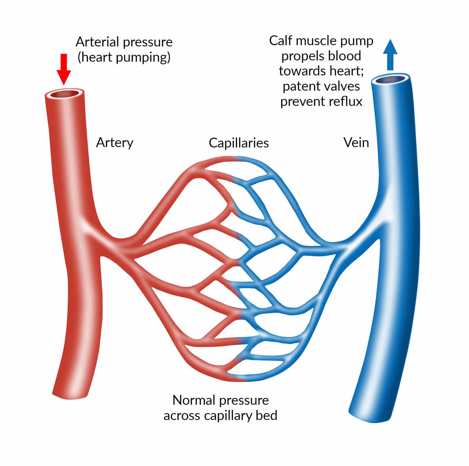
Blood is propelled through the arteries by pressure produced from the beating heart; that pressure is further maintained by the elastic nature of healthy arteries and their smaller branches (arterioles). Blood then passes through the capillary bed; a network of tiny capillaries within tissues (such as muscle and skin in the legs and feet). The capillaries have extremely thin walls (membranes) which allow oxygen and nutrients to pass into and supply the tissues. Blood returns to the heart via the low pressure veins with walls that are much thinner but tend to be larger in diameter than arteries.
This is why it is common to be able to see veins under the skin as the thin walls are more transparent to the blood they contain; it is rare to see an artery through the surface of the skin.
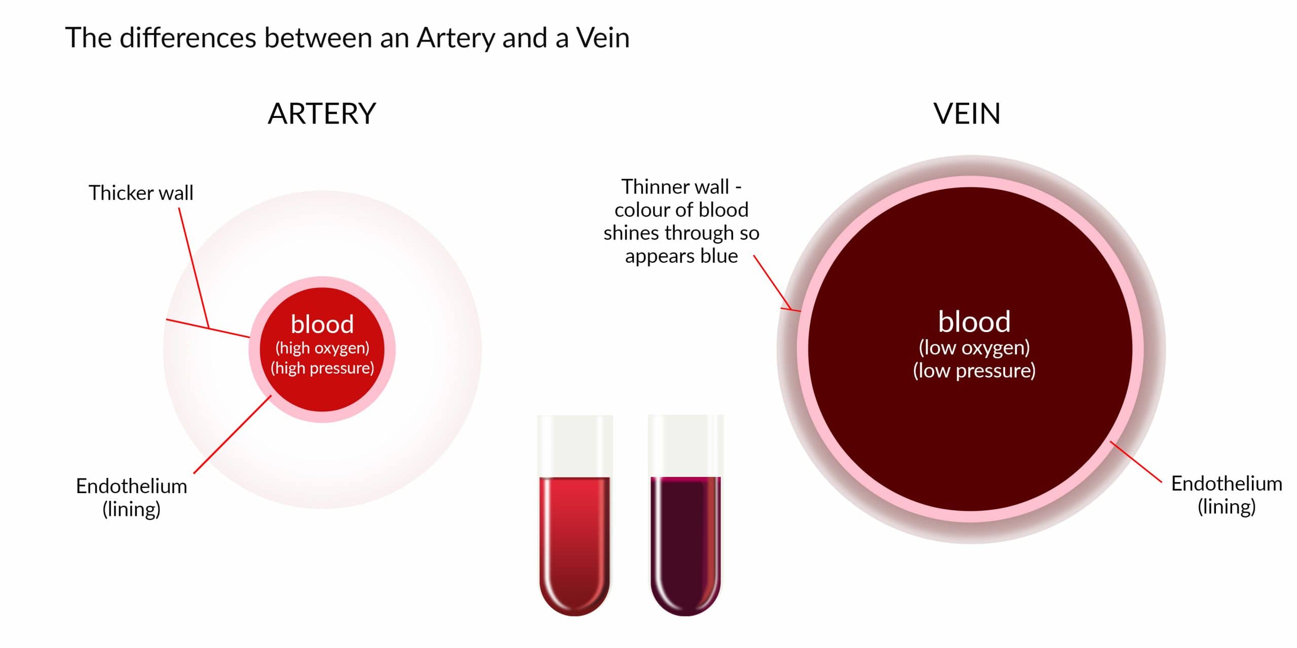
The colour of the blood varies with the amount of oxygen it contains. Oxygenated blood in arteries is bright red in colour whereas deoxygenated blood within the veins is a much darker and more blue colour. As a vascular surgeon I perform operations and procedures on both arteries and veins; I can identify the nature of an injured vessel by the colour of the blood.
Healthy leg veins
The contraction of muscles within the feet and the calves propel the blood back up through the veins towards the heart against gravity. A series of one-way valves allows the blood to travel in the correct direction.
The muscle compartments of the leg are surrounded by thick fibrous tissue called deep fascia. The venous system of the leg is broadly divided into two parts. The deep veins lie deep to this fascia and are in close proximity to the main arteries. The superficial veins are situated outside the deep fascia.
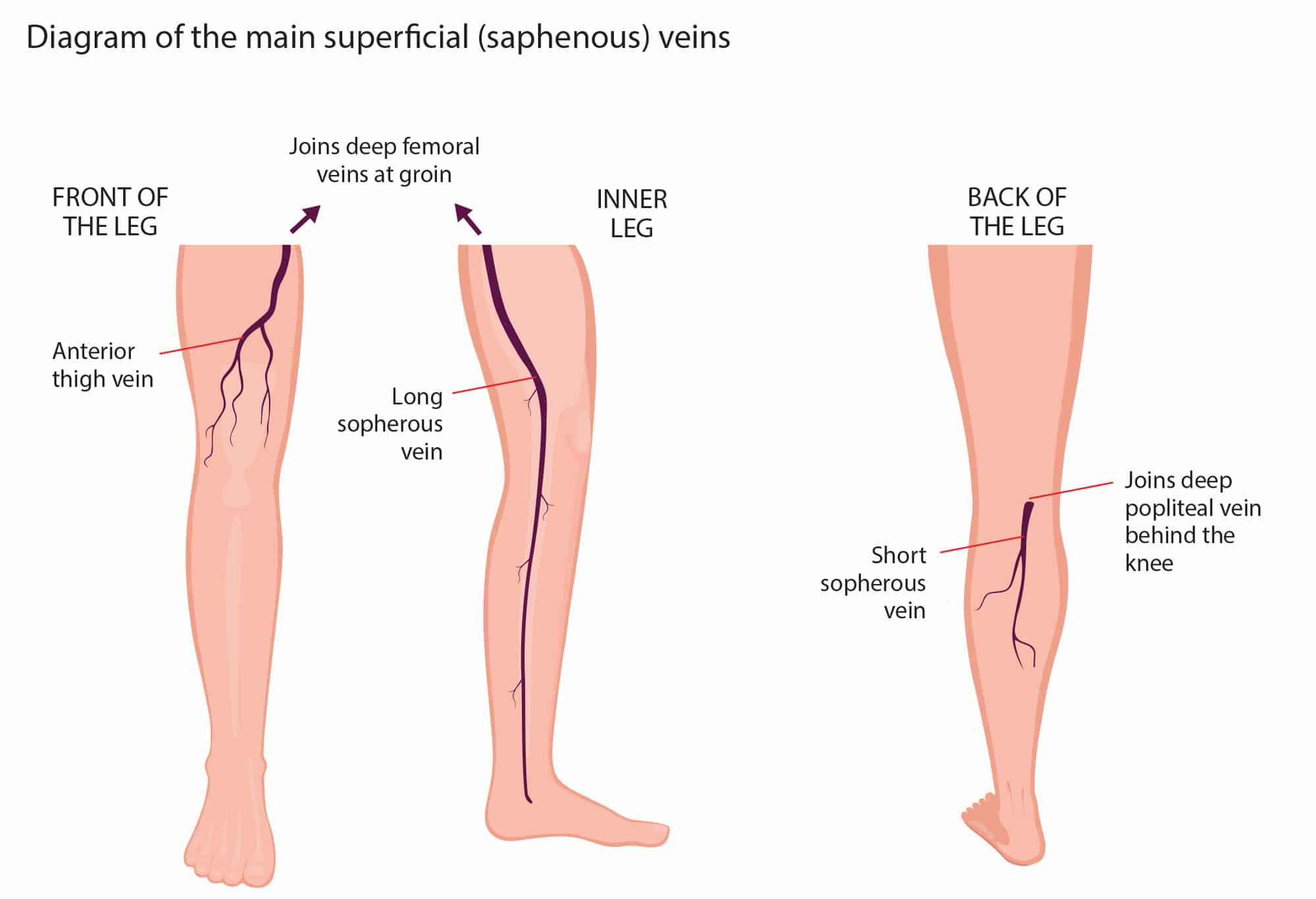
The long saphenous vein travels from the ankle up the inside of the calf and join the deep femoral veins at the groin.
The short saphenous vein travels from the outside of the ankle at the back of the calf and drain into the deep popliteal vein behind the knee.
The anterior thigh vein travels from the front and outside areas of the shin and thigh to drain into the deep femoral vessels at the groin.
FAQs…
Frequently Asked Questions
What are the main differences between normal veins and varicose veins?
Normal veins have smooth, elastic walls allowing them to efficiently return blood to the heart. They contain one-way valves ensuring blood flows in the correct direction. Varicose veins develop when these valves weaken or fail, causing blood to pool, which in turn makes the veins enlarge and twist, becoming more visible under the skin. The colour, bulging appearance, and associated discomfort or pain distinguish varicose veins from normal veins. They commonly occur in the legs and can cause aching, fatigue, and a heavy sensation in the affected limbs, among other symptoms.
What are the saphenous veins in the leg?
The saphenous veins in the leg are the great saphenous vein and the small saphenous vein. The great saphenous vein begins at the foot’s inner side and extends up the inner leg to the groin. The small saphenous vein originates on the outer side of the foot, runs up the calf’s back, and connects to the popliteal vein behind the knee. Both are superficial veins, lying close to the skin’s surface. They are essential for draining blood from the legs, channelling it back toward the heart. If dysfunctional, they can contribute to the development of varicose veins.
What is the difference between an artery and a vein?
Arteries and veins are both blood vessels but serve different functions. Arteries carry oxygenated blood from the heart to the rest of the body, with the exception of the pulmonary arteries, which carry deoxygenated blood to the lungs. They have thick, muscular walls to withstand the high pressure from the heart’s pumping. Veins, on the other hand, carry deoxygenated blood back to the heart from the body’s tissues, except for the pulmonary veins, which transport oxygenated blood from the lungs to the heart. Veins have thinner walls and contain one-way valves to prevent backflow of blood, especially in the limbs.
How can I tell if my veins are normal?
To determine if your veins are normal, observe their appearance and feel for any discomfort. Normal veins typically lie flat under the skin and are not overly visible unless you have thin or pale skin. They shouldn’t cause pain or discomfort. If veins become enlarged, twisted, bulging, or dark blue or purple, they may be varicose veins. If there’s swelling, warmth, redness, or tenderness around a vein, it could suggest a condition like thrombophlebitis.

Video appointments
Book your FREE initial consultation to discuss your varicose or thread vein removal via video. This enables us to meet, virtually, face to face and for us to diagnose your condition and answer any specific questions.